anatomy of the ear
We have already mentioned many things about how our ears work, and we have introduced some concepts related to the vibration of the eardrum and the perception of sound. In order to understand more complex measurements and sound behaviours, we need to take a closer look at our ears and know a little more about our hearing.
What we actually see of our hearing system is just what is called the tip of the iceberg: the external ear, or actually a part of it: the pinna or auricle.
The hearing system consists of three main parts:
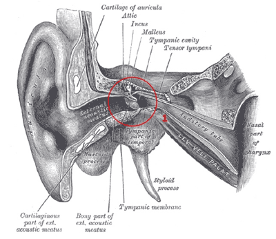
The auricle conveys the air into the external canal, at the end of which it hits the eardrum, a very thin membrane that collects the vibrations of the air particles (1). Attached to the eardrum there is a system of three little bones (not visible in first picture, it is located behind the eardrum).
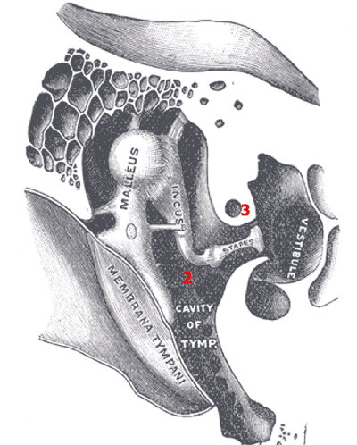
The malleus vibrates with the eardrum, and transfers its vibrations to the incus, which on its turn is connected to the stapes (2). The stapes, which is the smallest bone in the human body (approx. 0,2-0,3 cm long) conveys its vibrations to an oval window which is a membrane located on the cochlea. The cochlea is a spiral shaped organ filled with a liquid (Endolymph) that vibrates according to the movements of the oval window (3) (visible in picture 2 and 3).
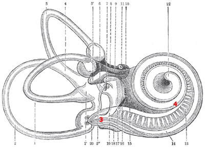
The internal walls of the cochlea are covered with a microscopic hairlike vibrating structure called cilia. The purpose of the cilia is to transform the movement of the fluid that surrounds them into electrical signals (4) that travel through the cochlea nerve to our brain. Each group of cilia only transduce a specific range of frequencies depending on their position in the cochlea. The closer they are to the oval window, the higher is the frequency that they transduce. It follows that the more distant ones will be deputed to transduce low frequencies.
The three loops on the right hand side of picture 3 are called 'semi-circular canals': they are filled of fluid which serve the purpose of maintaining our sense of balance, while the auditory tube visible in picture (1) connects the middle ear with the back of the nose: its purpose is to equalize the pressure in the middle ear according to the outer pressure.
Now that we have seen in a nutshell how we are able to hear, there are some facts that we must keep into account.
First of all, we must acknowledge that our ear has some physical limitations: due to the force necessary to move the eardrum membrane, we cannot hear every sound but only the ones that exercise a pressure of more than 20µPa. On the other hand, there is a limit the maximum pressure that our eardrum can handle: being a very thin membrane, it makes it subject to damages if exposed to too high pressure levels. Secondly, there is also a limit to the frequencies that we can hear: in the following lessons we are going to talk more in details about how our ear reacts to external stimuli.
What we actually see of our hearing system is just what is called the tip of the iceberg: the external ear, or actually a part of it: the pinna or auricle.
The hearing system consists of three main parts:
- the external ear, which is made by the pinna and the outer auditory canal
- the middle ear, which consists of the eardrum and of three bones called malleus, incus and stapes
- the inner ear, which is made by the cochlea and three semi-circular canals

The auricle conveys the air into the external canal, at the end of which it hits the eardrum, a very thin membrane that collects the vibrations of the air particles (1). Attached to the eardrum there is a system of three little bones (not visible in first picture, it is located behind the eardrum).

The malleus vibrates with the eardrum, and transfers its vibrations to the incus, which on its turn is connected to the stapes (2). The stapes, which is the smallest bone in the human body (approx. 0,2-0,3 cm long) conveys its vibrations to an oval window which is a membrane located on the cochlea. The cochlea is a spiral shaped organ filled with a liquid (Endolymph) that vibrates according to the movements of the oval window (3) (visible in picture 2 and 3).

The internal walls of the cochlea are covered with a microscopic hairlike vibrating structure called cilia. The purpose of the cilia is to transform the movement of the fluid that surrounds them into electrical signals (4) that travel through the cochlea nerve to our brain. Each group of cilia only transduce a specific range of frequencies depending on their position in the cochlea. The closer they are to the oval window, the higher is the frequency that they transduce. It follows that the more distant ones will be deputed to transduce low frequencies.
The three loops on the right hand side of picture 3 are called 'semi-circular canals': they are filled of fluid which serve the purpose of maintaining our sense of balance, while the auditory tube visible in picture (1) connects the middle ear with the back of the nose: its purpose is to equalize the pressure in the middle ear according to the outer pressure.
Now that we have seen in a nutshell how we are able to hear, there are some facts that we must keep into account.
First of all, we must acknowledge that our ear has some physical limitations: due to the force necessary to move the eardrum membrane, we cannot hear every sound but only the ones that exercise a pressure of more than 20µPa. On the other hand, there is a limit the maximum pressure that our eardrum can handle: being a very thin membrane, it makes it subject to damages if exposed to too high pressure levels. Secondly, there is also a limit to the frequencies that we can hear: in the following lessons we are going to talk more in details about how our ear reacts to external stimuli.
all images on this page taken from H. Grey's 'Anatomy of the Human Body', 20th edition, New York, Bartleby.com, 2000 courtesy and © Bartleby.com
